Our groups
The program Physics of Biology provides interdisciplinary training towards a doctorate (PhD) at the interface between Physics and Biology. Research in the program aims at understanding biological processes from physical principles using a large variety of different approaches. Groups participating in the program belong to various departments of the Faculties of Sciences and of Medicine. The goal of the program is to prepare you to pursue quantitative approaches to biological problems. It offers graduate-level coursework in physics and life sciences tailored to your background. By working on cutting-edge problems at the interface between biology and physics and by continuous participation in the lively seminar program in Geneva, you are led to develop your own independent research tracks.
Nonlinear nano- and bio-photonics
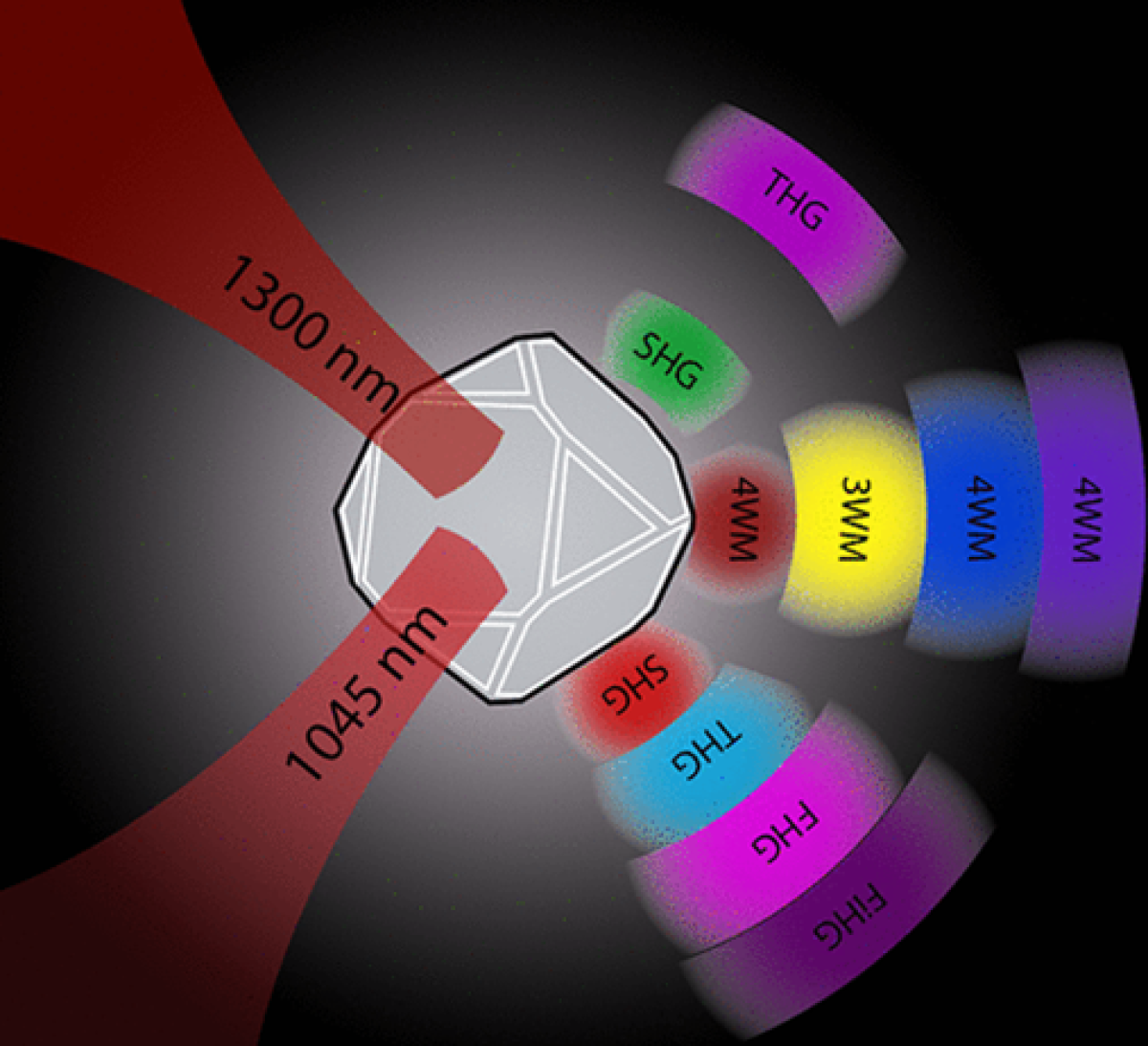
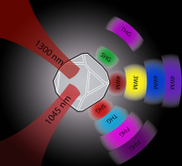
Light is a powerful tool for investigating living systems, starting from individual cells to tissues and whole organisms. Thanks to the increasing availability of new photonics tools, we can now widen the range of interactions of light with living matter and go beyond traditional fluorescence. In our group, we combine nonlinear optics and nanotechnology to provide bio-imaging devices with improved performances in terms of penetration depth, speed, selectivity and take part to interdisciplinary studies for the preclinical assessment of new diagnostics and therapeutic strategies.
Apply now
Physical principles of vertebrate regeneration
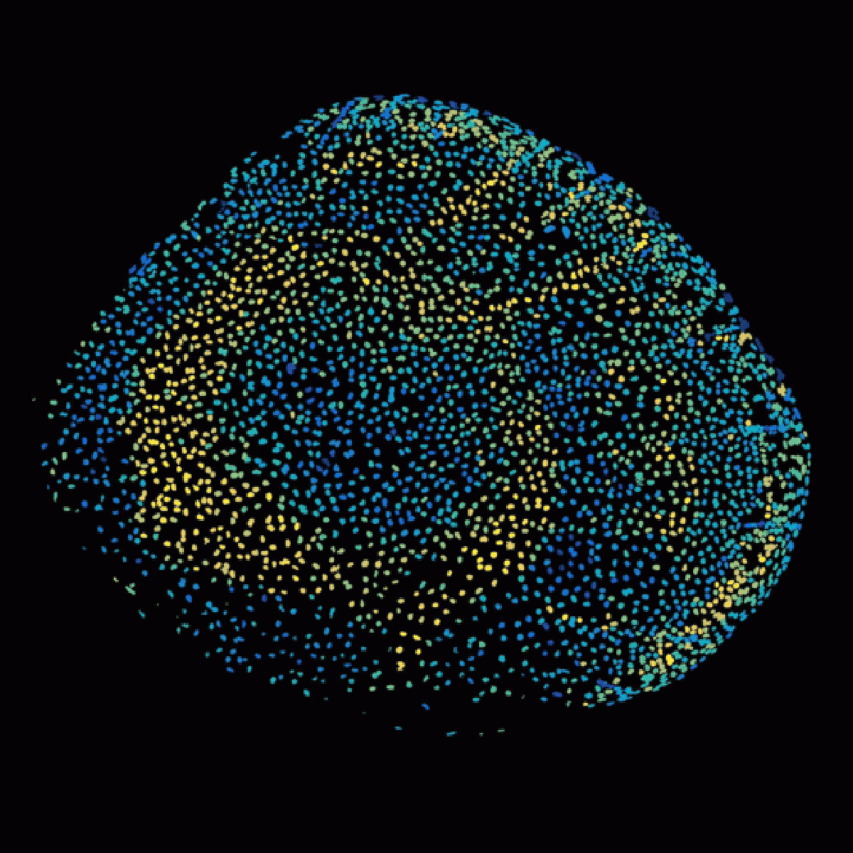
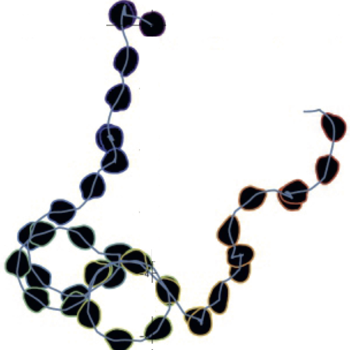
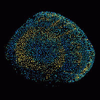
Regeneration is the astonishing process in which an animal regrows a lost body part. We are interested in how cells coordinate each other for a vertebrate body part to regenerate into its proper form. How are signals organized in time and space and integrated with tissue mechanics to orchestrate growth? This question lies at the boundary of physics and biology. To tackle it, we use experimental, computational, and theoretical methods and zebrafish as animal model. In particular, we like to use live imaging to quantify the signals, forces and tissue dynamics underlying regeneration of the zebrafish bony scale. We are looking for biologists, physicists, engineers, mathematicians (and more) who want to sail across disciplines in the search of the mechanisms of regeneration.
Apply now
Theoretical biological Physics
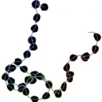
Our aim is to understand collective phenomena in living cells and other biological systems. To this end we use methods and concepts from theoretical physics to study specific systems preferentially in collaboration with experimental colleagues. From the point of view of physics, the challenge lies in treating systems out of thermodynamic equilibrium that have a biological function and an evolutionary history. To do justice to all of these aspects requires the development of new tools and approaches. Most of our projects address different aspects of cytoskeletal dynamics in the context of cell migration and division. In addition, we study pattern formation in bacteria, collective effects in cell signalling, and aspects of molecular evolution.
Apply now

Theoretical physics of biological development
We study how physical principles play a role during development of biological organisms. Properly orchestrated morphogenetic movements and tissue patterning allow animals to develop and establish their shape. We ask from a physical point of view how cell-cell communication and forces act together to shape the embryo. To reach that goal, we use methods from soft matter physics and non-linear dynamics and develop new theoretical and computational tools. Since biological systems function out-of-equilibrium, we also aim at understanding better the physical properties of active matter. We work in close collaboration with experimentalists to achieve a quantitative understanding of biological self-organization.
Apply now
Structural Systems Biology and Data Driven Biology
My lab will move to UniGE in March 2024. We explore principles by which proteins self-organize in cells. In this endeavor, we develop/use computational (AI, data science, image processing) and experimental (microscopy, synthetic biology, omics) approaches. We bridge scales from structure to phenotypes, with the goal of gaining a holistic understanding of life’s processes. Example of projects: (i) Physical and biological principles of protein phase-separation (collaboration with labs in NYC + Tokyo). (ii) Massively parallel prediction of protein structures and complexes with AI to analyze their evolution. (iii) Modeling and design of intrinsically disordered regions able to self-interact specifically.
Apply now
Medical Physics
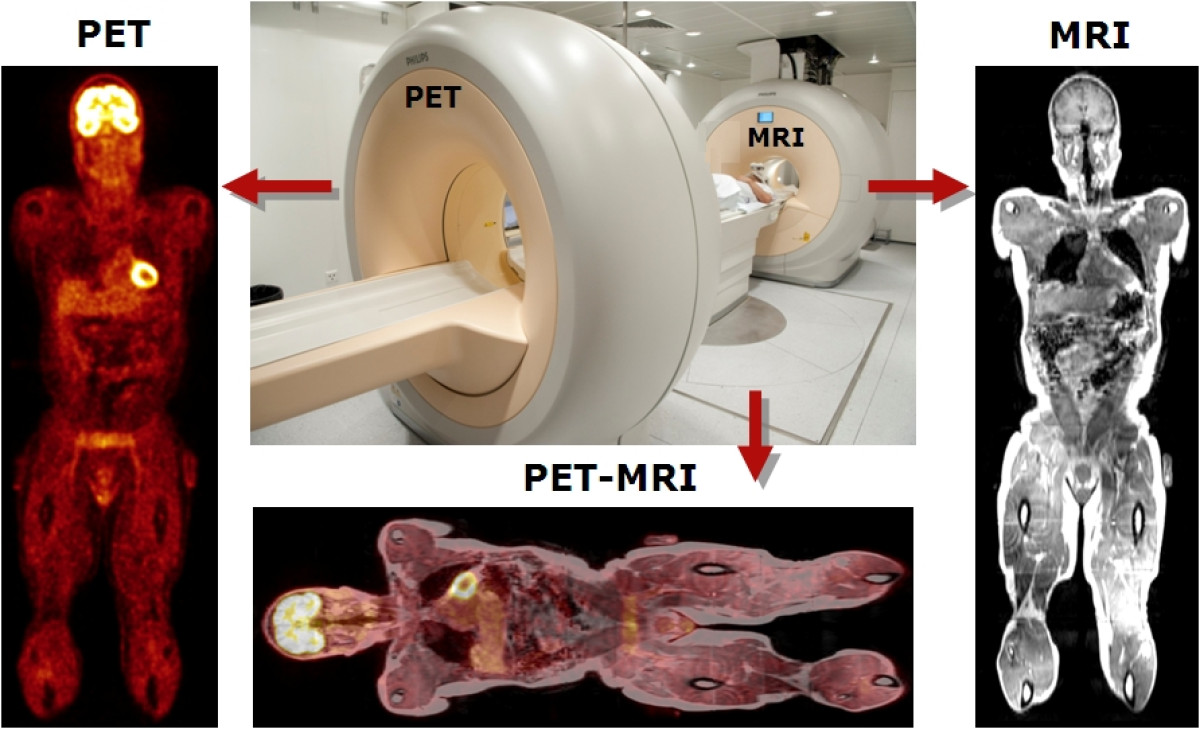
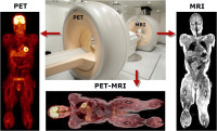
The PET Instrumentation & Neuroimaging laboratory (PinLab) has assumed a leading role and become internationally recognized for excellence in multimodality medical imaging research with PET being a focus for its activities. The group gained international recognition for contributions to the development and analysis of new image correction and reconstruction techniques for improved quantification of PET images as well as the development of computational models of human and animal models in connection with Monte Carlo-based dosimetry calculations. The lab has also been involved in the design and evaluation of hybrid imaging devices. PinLab provides an academic environment in a university hospital setting for training of highly qualified personnel in medical physics and multimodality imaging.
Apply now

Nanopore single-molecule detection of glycans
Glycans are major building blocks of life. They play a central role in many biological processes and are involved in most major diseases. However, glycoscience remains relatively understudied due to a lack of tools to probe the highly diverse, heterogeneous and complex structures of glycans. In contrast, the nanopore technique has been successfully applied to sequence long strand DNA, which has revolutionized the development of precision medicine. Therefore, we aim to develop biological nanopores as a novel analytical tool for glycan analysis, with the benefits of high sensitivity, low-cost, label-free, high-throughput, and potential for integrating into portable devices.
Apply now
Biophysics and Cell Biology of Signaling
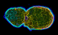
We want to understand in physical and molecular terms how cells talk to each other during development. This means our research is highly interdisciplinary: physics, cell biology, molecular biology, biochemistry, genetics... Indeed some of us in the lab are biologists, other physicists, chemists, engineers or mathematicians. We are interested in the signaling events that control tissue growth: how is the shape and final size of a tissue achieved during embryogenesis? We focus on two types of proliferation modes: growth control by morphogen gradients and asymmetric cell division in stem cells. We do this using two model systems: Drosophila and Zebrafish.
Apply now

Physics of Living Surfaces
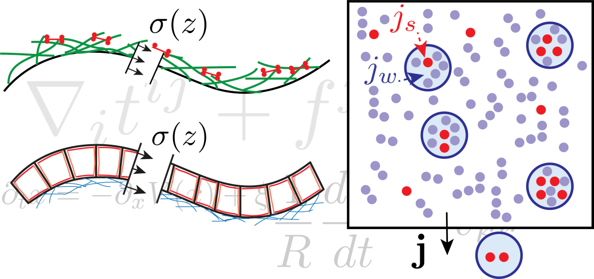
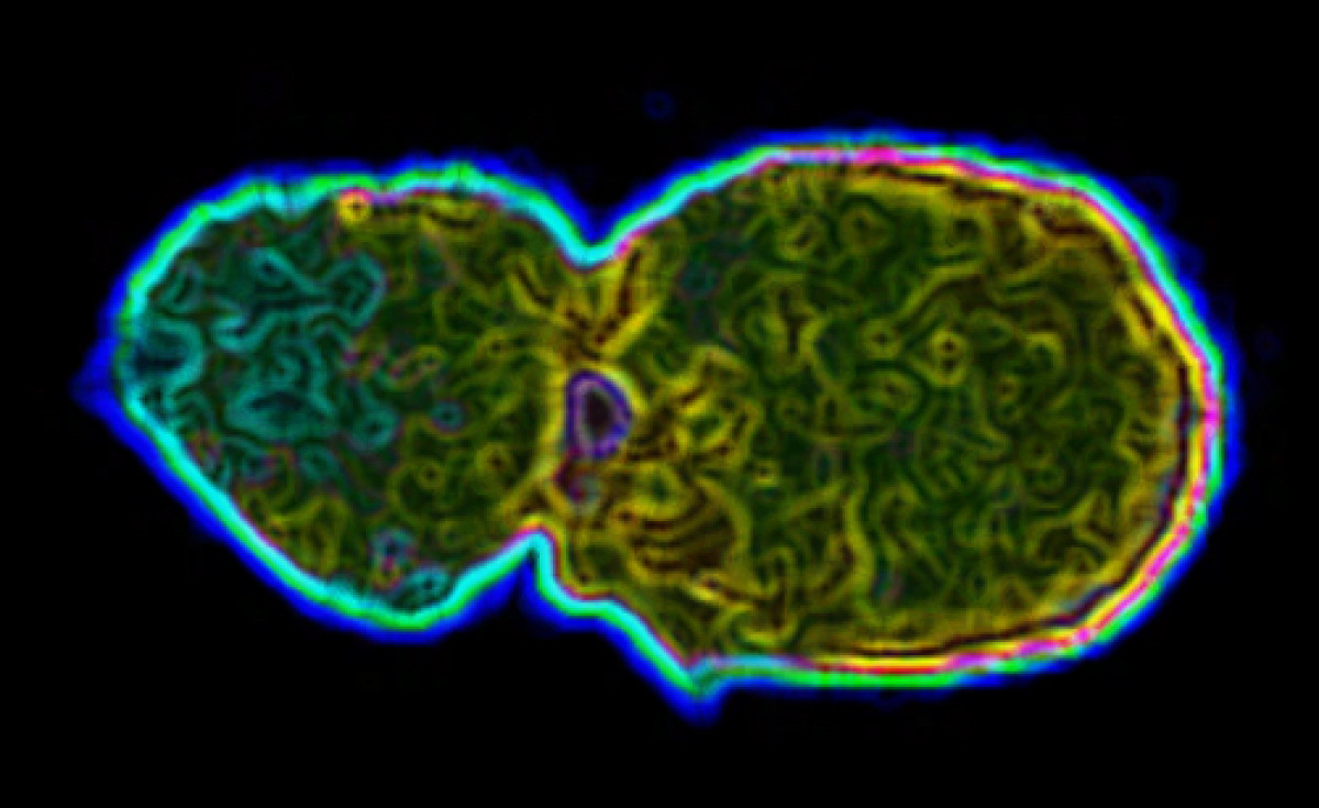
Many biological systems are made of viscoelastic surfaces. For example, the lipid bilayer that delimitates cells from their environment is a 2D fluid interface/barrier that can be deformed easily and is resistant to stretch. The plasma membrane is being constantly remodelled through processes such as endocytosis in membrane traffic. Another example, but at a different scale, is the epithelium, a cell monolayer that separates organs from the external environment. These epithelia grow and fold during development to form organs, being viscoelastic surfaces. We are interested in understanding how the unique physical properties of lipid membrane and epithelia respectively contribute to membrane traffic and organogenesis.
Apply now
Biochemistry and Biophysics of the Cytoskeleton
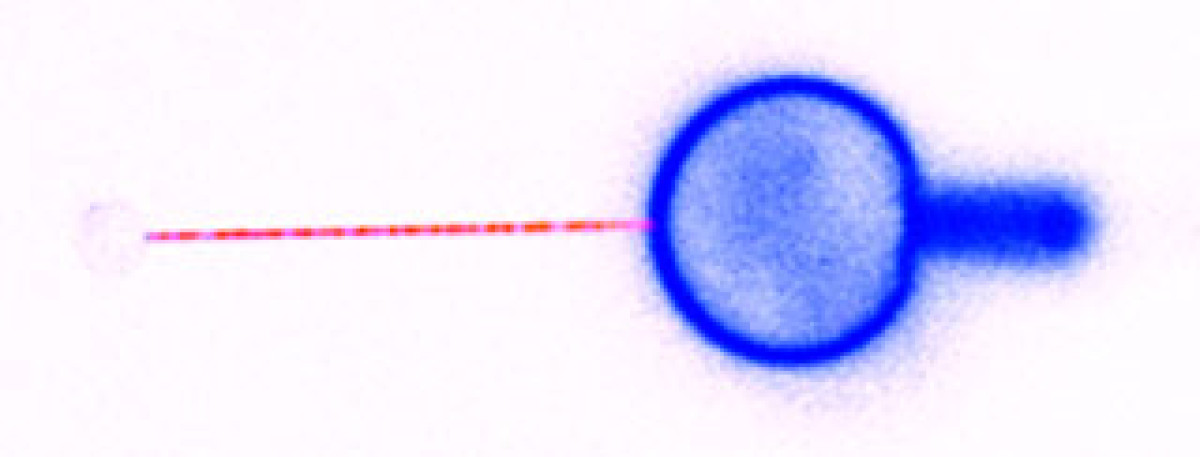
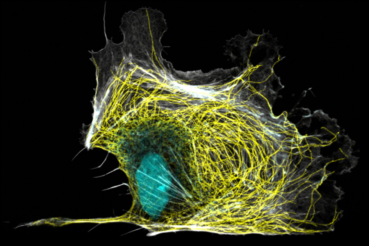
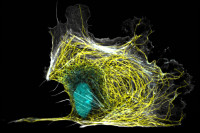
Cells have a complex actin and microtubule cytoskeleton network. This network is essential for many processes in cells: cell migration, division, differentiation…. We want to understand how the cytoskeleton is organized and regulated in space and time, resulting in a functional architecture. To understand the underlying principles, we study the cytoskeleton dynamics, mechanics and interactions using two systems: cell lines and isolated proteins.
Apply now
Biomedical Engineering and Interventional Imaging
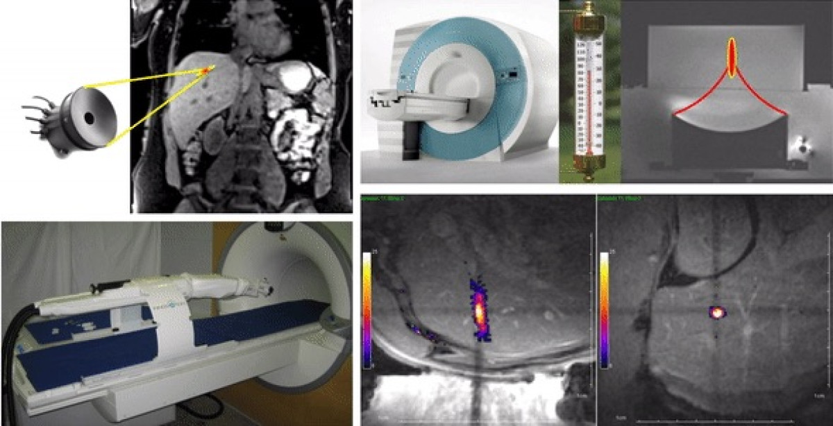
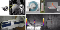
The work group comprises research scientists and radiologists. The team has a wide range of competences in biomedical engineering, interventional radiology, intra-operative image guidance, computer sciences. Our topics cover translational and clinical research, in particular in the field of oncology, from in-house prototyping of innovative devices to clinical trials using image-guided novel therapies. High intensity focused ultrasound (HIFU) is currently the only extra-corporeal technology capable of depositing sharply delineated energy patterns deep inside the body. Magnetic resonance imaging provides excellent capabilities for spatial guidance of HIFU therapy and is currently the only available technique for non-invasive temperature measurement within biological tissue.
Apply now
Patterning of the vertebrate skin through mechanical and Turing instabilities
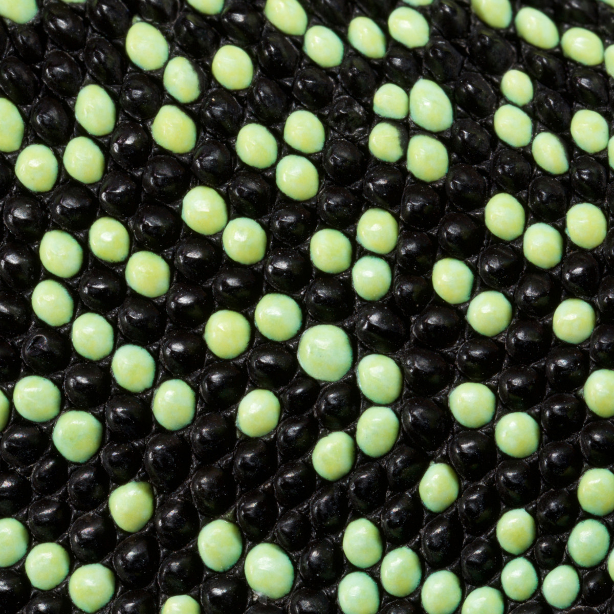
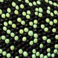
Because discipline-oriented approaches can sometimes restrain imagination, our laboratory includes biologists, physicists and computer scientists, making our research highly multidisciplinary and integrative. One of our main models is the development and evolution of the vertebrate skin, with special emphasis on skin appendages (scales, hairs, spines), skin colours (pigmentary and structural), and skin colour patterns. Our strategy is to tackle these topics from different angles by using multiple techniques (genomics, physical experiments, mathematical modelling and numerical simulations). We work at multiple spatial scales (genomes, cells, tissues, organisms) and use new model species of tetrapods (frogs, snakes, lizards, crocodiles, hedgehogs, tenrecs, spiny mice).
Apply now
